FRCR Part 2A · Genitourinary · Testicular pathology
Testicular Pathology for FRCR Part 2A
Published by FRCR AI · AI-assisted educational content
Created and overseen by Dr. Tan Min On, Radiologist — MedNex Pte. Ltd.
Published · Updated
Essential Testicular Pathology for the FRCR Part 2A Exam
Mastering the current WHO 5th Edition (2022) classification and AJCC 8th edition staging is essential for clinical success and board examination readiness in genitourinary radiology. This overview covers the critical diagnostic criteria and staging paradigms required for the FRCR Part 2A.
1. The Primacy of GCNIS and Germ Cell Tumors
Germ cell neoplasia in situ (GCNIS) is the established precursor to the vast majority of testicular germ cell tumors (TGCTs). Clinically, all solid testicular masses must be managed as malignant until proven otherwise, usually necessitating surgical exploration. Germ cell tumors are primarily divided into Seminomatous (SGCT) and Non-seminomatous (NSGCT) types, with distinct prognostic and therapeutic pathways governed by histopathology and serum tumor markers. Exam tip: Always correlate imaging findings with AFP, hCG, and LDH levels. Crucially, an elevated AFP level effectively rules out a diagnosis of pure seminoma and confirms the presence of non-seminomatous elements.
2. AJCC 8th Edition Staging Nuances
The AJCC 8th edition introduced specific changes regarding seminoma staging that are frequent targets for FRCR questions. Specifically, pathological T1 (pT1) is now subdivided based on tumor size: pT1a refers to a tumor less than 3 cm in greatest dimension, while pT1b refers to a tumor 3 cm or greater in greatest dimension. Exam tip: Remember that this size-based pT1 sub-classification applies exclusively to pure seminomas. It is not applied to NSGCTs.
3. Updated WHO 2022 Terminology
Updates to the WHO classification include replacing deprecated terms to improve diagnostic precision and align with international standards. "Carcinoid" is now formally termed "neuroendocrine tumor (NET)," and the entity previously known as "well-differentiated papillary mesothelioma" is now "well-differentiated papillary mesothelial tumor," reflecting its generally favorable clinical behavior. Furthermore, the term "teratoma with primitive neuroectodermal tumor (PNET)" has been updated to "teratoma with embryonic type neuroendocrine differentiation." Exam tip: Avoid using legacy terms like carcinoid or PNET on written exams. Using the current WHO 2022 nomenclature is vital for high-scoring responses.
4. Sonographic Differential Diagnosis
Ultrasound remains the primary imaging modality for scrotal pathology. While malignancy remains the priority, benign conditions can mimic tumors. The following table summarizes key comparative features.
| Condition | Imaging Features | Key Association |
|---|---|---|
| Seminoma | Hypoechoic, homogenous | Most common pure GCT |
| Teratoma | Heterogeneous, calcification | Common in pre-pubertal age group |
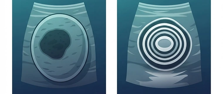
| Epidermoid Cyst | Onion-skin pattern | Benign, avascular, target-like |
| Lymphoma | Diffuse enlargement, hypoechoic | Common in patients > 60 years |
Exam tip: An avascular, concentric 'onion-skin' mass is the pathognomonic description for a testicular epidermoid cyst. Recognizing this benign entity is essential to avoid unnecessary radical orchiectomy.
5. Standardizing Pathologic Reporting
Modern oncological pathology has moved away from reporting mitotic activity per high-power field (HPF), as the field of view varies significantly between microscope models. Standardization now mandates reporting mitotic counts per mm² to ensure reproducibility and prognostic accuracy. Exam tip: If a question mentions mitotic counts, recognize that standardized area-based reporting is the current professional requirement in pathology.
6. Emerging Entities
Candidates should be aware that the 2022 WHO classification has introduced new entities to the nomenclature, including the signet ring stromal tumor, the myoid gonadal stromal tumor, and the pre-pubertal type testicular neuroendocrine tumor. While rare, their inclusion in the latest guidelines highlights the shift toward more granular pathological assessment.
Key Takeaway: The FRCR exam prioritizes the application of the WHO 2022 classification and the AJCC 8th edition staging. Focus on the size-based pT1 sub-classification for seminomas, recognize GCNIS as the foundational precursor, and utilize current terminology to demonstrate an up-to-date understanding of genitourinary malignancy.
Topics: testicular ultrasound · frcr part 2a · genitourinary radiology · germ cell tumors · who classification