FRCR Part 2A · Paediatric · Developmental anomalies
Paediatric Developmental Anomalies: High-Yield FRCR Part 2A Pearls
Published by FRCR AI · AI-assisted educational content
Created and overseen by Dr. Tan Min On, Radiologist — MedNex Pte. Ltd.
Published · Updated
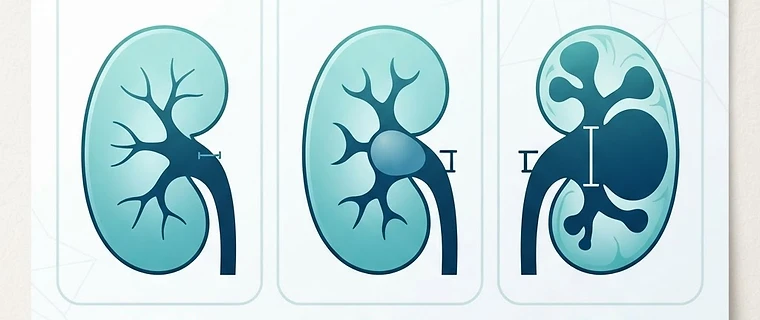
High-Yield Concepts in Paediatric Developmental Anomalies
Mastering paediatric developmental anomalies requires familiarity with updated international classifications and a shift toward genotypic-phenotypic integration. This guide provides high-yield pearls designed for the FRCR Part 2A examination, reflecting the most recent consensus guidelines.
1. CPAM and Pulmonary Malformations
Congenital Pulmonary Airway Malformations (CPAM) represent a spectrum of lung lesions, replacing the outdated term CCAM. It is critical to differentiate CPAM from bronchopulmonary sequestration (BPS) on cross-sectional imaging; BPS is defined by a systemic arterial supply, typically from the aorta. Exam tip: Always look for a systemic feeder vessel on CT or MRA when evaluating a cystic pulmonary lesion in an infant. Remember that CPAMs communicate with the tracheobronchial tree, whereas BPS typically does not.
2. Standardising Hydronephrosis: The UTD System
Subjective terms such as 'mild', 'moderate', or 'severe' hydronephrosis are deprecated in modern reporting, having been replaced by the Urinary Tract Dilatation (UTD) classification. This system uses a combination of AP renal pelvis diameter, calyceal dilation, and parenchymal thickness to provide an objective risk-stratification score. This classification is now central to the evaluation of Congenital Anomalies of the Kidney and Urinary Tract (CAKUT).
| UTD Category | Risk Stratification | Criteria Summary |
|---|---|---|
| UTD P1 | Low risk | Normal parenchymal thickness, minimal pelvic dilation |
| UTD P2 | Intermediate | Pelvic dilation with minor calyceal or parenchymal changes |
| UTD P3 | High risk | Significant pelvic/calyceal dilation, parenchymal thinning |
Exam tip: Memorise the UTD classification components; examiners focus on the objective criteria rather than subjective qualitative descriptions.
3. Vascular Anomalies: The ISSVA Framework
The International Society for the Study of Vascular Anomalies (ISSVA) classification is the global standard, strictly separating vascular malformations from vascular neoplasms. Vascular malformations are structural errors of vasculogenesis (low or high flow, present at birth), whereas vascular neoplasms like infantile haemangioma undergo rapid proliferation followed by involution. Exam tip: Infantile haemangiomas typically demonstrate intense, homogeneous enhancement on MRI and exhibit flow-voids as they transition into the involution phase. Conversely, vascular malformations are often stable in size.
4. Skeletal Dysplasia and Gene-Linked Naming
The 11th revision of the International Skeletal Dysplasia Society (ISDS) nosology has moved away from eponymous naming (e.g., Fairbank disease) toward a dyadic system linking phenotypes to their causative genes (e.g., COL2A1-related disorders). When describing skeletal dysplasias, focus on the predominant distribution of abnormalities: rhizomelic (proximal), mesomelic (middle), or acromelic (distal). Exam tip: Rhizomelic shortening points toward classic achondroplasia, while acromelic involvement is more characteristic of chondroectodermal dysplasia. Be prepared to identify the gene-phenotype association.
5. Potter Sequence: More Than Just Renal Agenesis
Potter Sequence is the preferred term over 'syndrome', as it describes the cascading phenotype resulting from severe oligohydramnios. While bilateral renal agenesis is the classic cause, posterior urethral valves (PUV) and chronic amniotic fluid leakage are frequent triggers for the same morphological consequences (pulmonary hypoplasia and abnormal facies). Exam tip: Identify the 'keyhole sign' on ultrasound as the pathognomonic finding for posterior urethral valves in a male neonate with suspected Potter sequence. Additionally, recall that fetal MRI is increasingly used to quantify lung volumes to aid prognosis.
6. Müllerian and CNS Updates
When assessing female pelvic anomalies, utilise the 2021 ASRM classification (9 categories) rather than older AFS classes. In CNS imaging, remember the 2021 WHO 5th Edition classification updates, which mandate the use of the term 'type' and 'subtype' over 'variant', and ensure that WHO grading is always represented by Arabic numerals (e.g., IDH-mutant astrocytoma, CNS WHO grade 2) rather than Roman numerals.
Key Takeaway: Success in the FRCR Part 2A requires strict adherence to current nomenclature, including the use of CPAM, UTD grading, and the ISSVA classification, while moving away from outdated eponymous terms in skeletal and oncological radiology.
Topics: paediatric radiology · frcr 2a · developmental anomalies · fetal mri · utd classification