FRCR Part 1 Physics · Nuclear Medicine · Internal dosimetry
Internal Dosimetry in Nuclear Medicine
Published by FRCR AI · AI-assisted educational content
Created and overseen by Dr. Tan Min On, Radiologist — MedNex Pte. Ltd.
Published · Updated
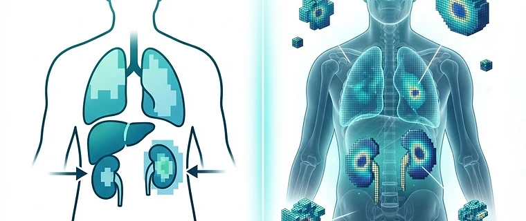
Internal Dosimetry in Nuclear Medicine: A Modern Framework
Modern internal dosimetry has transitioned from generalized, population-based estimates to a sophisticated, patient-specific discipline defined by the shift toward precision theragnostics. This guide covers the essential principles for FRCR Part 1 physics, emphasizing the updated terminology found in ICRU Report 96 and EANM clinical guidelines.
1. The Evolving MIRD Schema
The MIRD framework remains the fundamental equation: Mean Absorbed Dose (D) equals the product of Time-Integrated Activity (TIA) and the Dose Factor (S-value). While the traditional mathematical structure holds, we have shifted from 'Cumulated Activity' to the officially recognized 'Time-Integrated Activity (TIA)' and 'Time-Integrated Activity Coefficient' (formerly residence time). These refinements ensure greater harmonization in reporting across international clinical trials. Exam tip: Know the MIRD equation components: D = Ã × S, where à is the Time-Integrated Activity.
2. Transitioning to Patient-Specific Models
We are moving away from stylized hermaphroditic phantoms, which utilized averaged anatomical assumptions, toward voxel-based and patient-specific models. Modern dosimetry utilizes Monte Carlo simulations or Dose Voxel Kernels (DVK) derived directly from patient SPECT/CT or PET/CT datasets to account for individual organ shape and density. This precision is essential for avoiding nephrotoxicity in ¹⁷⁷Lu-based therapies. Exam tip: Recognize that voxel-based dosimetry allows for 3D dose distribution mapping, accounting for inhomogeneous activity distribution, unlike earlier 2D geometric averages.
3. Therapeutic Window and BED
In therapeutic applications, particularly with ¹⁷⁷Lu-labeled compounds, the objective is to optimize the therapeutic window while avoiding dose-limiting toxicities in critical organs like the kidneys and bone marrow. We now apply the Biologically Effective Dose (BED) model, imported from external beam radiotherapy, which accounts for the biological repair of low-dose-rate radiation exposure and the LQ (Linear-Quadratic) model of cellular survival. Exam tip: Remember that BED adjusts for the 'sparing effect' of protracted radiation delivery inherent in radionuclide therapy compared to high-dose-rate external beam therapy.
4. Alpha-Particle Dosimetry
The emergence of targeted alpha therapy (e.g., ²²⁵Ac) necessitates micro-dosimetry due to the high-LET and extremely short path length of alpha emissions. Unlike beta particles, which deposit energy over millimeters, alpha particles deposit energy over micrometers, requiring advanced modeling to assess damage at the cellular level. This requires careful consideration of the Relative Biological Effectiveness (RBE) and the localization of the radionuclide relative to the cell nucleus. Exam tip: Alpha-emitters have high RBE; dosimetry focuses on microscopic energy deposition patterns rather than organ-level mean absorbed dose alone.
| Concept | Old Term | New/Current Term |
|---|---|---|
| Metric | Cumulated Activity | Time-Integrated Activity (TIA) |
| Parameter | Residence Time | Time-Integrated Activity Coefficient |
| Phantom | Stylized Phantom | Voxel-based Patient-specific Model |
5. AI Integration in Dosimetry
Recent technological leaps involve using deep learning for the automated segmentation of organs at risk (OARs). By automating the contouring process, AI significantly reduces the time required to generate rapid, personalized dose maps, facilitating real-time clinical decisions during multi-fraction therapeutic regimens. This is crucial as clinical dosimetry becomes a standard of care rather than a research-only endeavor. Exam tip: AI is primarily used to overcome the bottleneck of manual voxel-based segmentation in clinical workflows.
Key Takeaway: Internal dosimetry has evolved from generalized models to a personalized, voxel-based approach using the ICRU 96 framework, incorporating BED for low-dose-rate radiobiology and AI for efficient clinical implementation.
Topics: nuclear medicine · physics · frcr · dosimetry · theragnostics