FRCR Part 2A · Neuroradiology · Stroke
Hyperacute Stroke Imaging and Management: FRCR Part 2A Pearls
Published by FRCR AI · AI-assisted educational content
Created and overseen by Dr. Tan Min On, Radiologist — MedNex Pte. Ltd.
Published · Updated
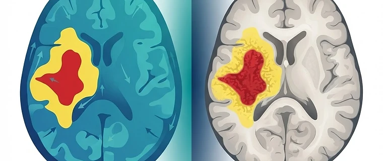
Hyperacute Stroke Imaging: Moving Beyond the Clock
Contemporary management of acute cerebrovascular syndromes has shifted from rigid time-based criteria to physiology-driven patient selection, leveraging advanced neuroimaging to define the salvageable penumbra. Understanding these paradigms is essential for the FRCR Part 2A examination.
1. The Decline of the '4.5-Hour Wall'
Clinical decision-making now prioritizes imaging-based triage over time-from-onset. While Alteplase and Tenecteplase (TNK) remain standard within 4.5 hours, perfusion mismatch (based on WAKE-UP or EXTEND criteria) allows for intravenous thrombolysis up to 9 hours in specific cohorts. Mechanical thrombectomy (EVT) is now indicated up to 24 hours in cases of clinical-imaging mismatch, as established by the DAWN and DEFUSE 3 trials. Exam tip: Always look for 'penumbral mismatch' on CT Perfusion (CTP) or MRI perfusion maps—where the volume of tissue at risk (penumbra) significantly exceeds the volume of the established infarct core.
2. Evolving ASPECTS and Core Assessment
The Alberta Stroke Program Early CT Score (ASPECTS) remains the cornerstone of MCA-territory assessment, though its application has expanded. Current literature, supported by trials such as SELECT2 and ANGEL-ASPECT, now explicitly includes patients with large-core infarcts (ASPECTS 3–5) for EVT, moving away from previous strict cut-offs. Furthermore, specialized scoring systems like pc-ASPECTS (posterior circulation) and AC-ASPECTS (anterior cerebral) are increasingly utilized to guide management in non-MCA territories. Exam tip: Automated AI-driven e-ASPECTS is now a standard clinical tool. However, candidates must be proficient in manual 10-point scoring as this remains the gold standard for clinical validation.
3. mTICI and Reperfusion Success
The modified Thrombolysis in Cerebral Infarction (mTICI) scale is the current standard for quantifying recanalization success. The modern definition distinguishes 2b as >50% reperfusion, while 2c (90-99%) is utilized to categorize 'near-complete' reperfusion, reflecting superior clinical outcomes.
| Score | Definition | Clinical Implication |
|---|---|---|
| 0 | No perfusion | Complete occlusion |
| 1 | Antegrade reperfusion past occlusion, distal branches limited | Minimal reperfusion |
| 2a | Reperfusion of <50% of target territory | Partial reperfusion |
| 2b | Reperfusion of >50% of target territory | Substantial reperfusion |
| 2c | Reperfusion of 90-99% of target territory | Near-complete recanalization |
| 3 | Complete reperfusion | Full vessel patency |
Exam tip: Focus on the distinction between 2b and 2c for MCQs regarding post-intervention success markers.
4. Expanding the Scope of EVT and DMVO
Mechanical thrombectomy is no longer restricted to proximal Large Vessel Occlusions (LVOs) of the ICA or M1 segment. Growing evidence supports the use of EVT for Distal Medium Vessel Occlusions (DMVOs), including M2/M3 and A2/A3 segments, particularly when clinical deficits are disabling. AI-based triage for these vessels is now standard in many hyperacute pathways. Exam tip: Do not reflexively exclude patients based on distal occlusions if the clinical syndrome is severe and the imaging suggests salvageable tissue.
5. Pharmacological Updates: Tenecteplase
Tenecteplase (TNK) is now widely endorsed as an alternative, and often preferred, option to Alteplase for bridging therapy. Its single-bolus administration offers superior logistical convenience in the hyperacute setting, particularly for patients requiring urgent transfer for EVT. Exam tip: If a clinical scenario involves complex logistics or transfer to a comprehensive stroke center, TNK’s single-bolus nature is a distinct advantage compared to the 1-hour infusion required for Alteplase.
Key Takeaway: Modern stroke imaging centers on penumbral identification rather than time-stamps. Prioritize the assessment of mismatch, acknowledge the inclusion of large-core infarcts, and master the updated mTICI scale to report post-intervention success accurately.
Topics: stroke · ct perfusion · thrombectomy · neuroradiology · frcr