FRCR Part 1 Physics · Nuclear Medicine · Gamma camera
Gamma Camera Systems and Molecular Imaging Physics
Published by FRCR AI · AI-assisted educational content
Created and overseen by Dr. Tan Min On, Radiologist — MedNex Pte. Ltd.
Published · Updated
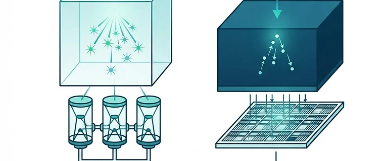
Gamma Camera Systems and Molecular Imaging Physics
Modern nuclear medicine has transitioned from legacy analog systems to sophisticated quantitative molecular imaging platforms, necessitating a firm grasp of both detector physics and hybrid anatomical integration. This summary focuses on the high-yield technical principles essential for the FRCR Part 1 Physics examination, aligning with current clinical standards such as AAPM Report No. 177.
1. Detector Evolution: NaI(Tl) to CZT
Traditional gamma cameras utilize Sodium Iodide (NaI) scintillation crystals coupled to photomultiplier tubes (PMTs). Newer solid-state Cadmium Zinc Telluride (CZT) detectors represent a paradigm shift by converting gamma photons directly into electrical signals without an intermediate light conversion step, offering superior energy resolution and spatial precision. Unlike legacy systems, these detectors are pixelated, allowing for compact, modular designs that support high-resolution imaging. *Exam tip: CZT detectors provide significantly better energy resolution than NaI crystals, which drastically improves scatter rejection and enables simultaneous multi-isotope imaging, a core capability in modern molecular imaging.
2. Energy Discrimination and Scatter Rejection
To maintain high signal-to-noise ratios, gamma cameras employ a pulse height analyzer (PHA). By setting a 20% acceptance window centered on the photopeak of the radionuclide, the system effectively rejects low-energy photons that have undergone Compton scattering within the patient. While this remains the standard for general applications, digital detector systems now allow for more precise multi-peak windows to handle complex isotopes like ¹¹¹In or ¹⁷⁷Lu with greater sensitivity. *Exam tip: Know that the 20% energy window is a standard compromise between sensitivity (total counts) and specificity (scatter rejection). Modern digital detectors enhance this by minimizing baseline drift common in older analog circuitry.
3. The Role of Collimators
Collimators are the primary determinants of the trade-off between spatial resolution and sensitivity in gamma camera systems. Parallel-hole collimators remain the clinical standard, but the choice of aperture size and bore length directly dictates the resulting image quality.
| Collimator Type | Sensitivity | Spatial Resolution | Typical Use |
|---|---|---|---|
| Low Energy High Resolution | Low | High | Thyroid, Bone Scan |
| Low Energy All Purpose | Medium | Medium | General SPECT |
| Low Energy High Sensitivity | High | Low | Cardiac First Pass |
*Exam tip: Remember the fundamental trade-off: increasing collimator hole length improves spatial resolution but decreases sensitivity due to the reduction in the solid angle of accepted photons.
4. Hybrid Imaging and Quantitative Standards
Standalone SPECT systems are increasingly considered deprecated in clinical practice. The industry standard is now hybrid SPECT/CT, which is essential for providing accurate attenuation correction and anatomical localization. Furthermore, the field is shifting from qualitative “hot spot” analysis toward Quantitative SPECT, which calculates absolute activity concentration (Bq/mL). This transition is vital for accurate dosimetry in theranostics. *Exam tip: AAPM Report No. 177 is the primary clinical benchmark for acceptance testing and annual surveys of these integrated systems. Be prepared to discuss how attenuation correction is fundamentally linked to the CT component of the hybrid unit.
5. Digital Detectors and Silicon Photomultipliers (SiPMs)
The move away from bulky vacuum-tube PMTs toward digital SiPMs allows for more compact, modular detector geometries. These digital architectures support faster count rates and improved timing resolution, which are critical for the increasing demands of modern AI-enhanced reconstruction algorithms. Digital detector arrays also exhibit greater immunity to external electromagnetic interference, enhancing the reliability of signal acquisition in clinical environments.
Key Takeaway: Success in the FRCR Physics exam requires moving beyond analog concepts toward an understanding of direct-conversion detectors (CZT), the necessity of hybrid SPECT/CT for quantitative accuracy, and the strict relationship between collimator geometry and image sensitivity. Ensure your knowledge reflects the modern shift toward molecular imaging as a quantitative, rather than purely qualitative, discipline.
Topics: nuclear medicine · gamma camera · physics · spect · molecular imaging