FRCR Part 1 Physics · X-ray Imaging · Image quality
FRCR Part 1 Physics: X-Ray Image Quality
Published by FRCR AI · AI-assisted educational content
Created and overseen by Dr. Tan Min On, Radiologist — MedNex Pte. Ltd.
Published · Updated
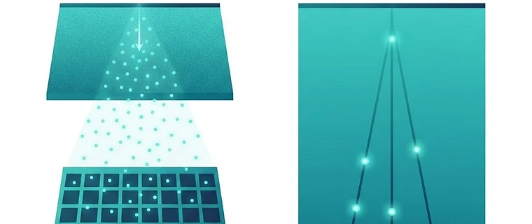
Mastering Image Quality in Digital X-Ray Imaging
Understanding image quality in modern digital radiology is critical for FRCR Part 1 success, focusing on objective metrics that define system performance and dose efficiency in the era of flat-panel detectors. Modern digital imaging prioritizes the optimization of diagnostic detail relative to radiation dose within the framework of IR(ME)R 2017 regulations.
1. The Objective Metrics of System Performance
Image quality is defined by three primary quantitative metrics: Modulation Transfer Function (MTF), Noise Power Spectrum (NPS), and Detective Quantum Efficiency (DQE).
- Modulation Transfer Function (MTF): Describes the ability of the system to transfer contrast from the object to the image at various spatial frequencies. It is effectively the spatial frequency response of the imaging system.
- Noise Power Spectrum (NPS): Characterizes the noise texture of the system, quantifying how noise power is distributed across different spatial frequencies.
- Detective Quantum Efficiency (DQE): The overarching metric of dose efficiency. It represents the efficiency of the detector in converting x-ray photons into a high-quality diagnostic image compared to an ideal detector. A higher DQE means the system can produce the same image quality at a lower radiation dose, which is a fundamental requirement of dose optimization under IR(ME)R.
2. Standardized Dose Indicators
With the shift to standardized digital systems, manufacturers have moved away from proprietary exposure indices (like S-numbers). The IEC 62494-1 standard mandates the use of Exposure Index (EI), Target Exposure Index (EIT), and Deviation Index (DI).
- Deviation Index (DI): Provides a direct numerical value representing how far the actual exposure is from the target. A DI of 0 indicates that the target exposure was achieved.
- Clinical Application: A negative DI indicates underexposure, increasing the risk of quantum mottle and loss of diagnostic information. A positive DI suggests overexposure, which results in unnecessary patient dose and potential 'exposure creep,' where clinicians inadvertently increase doses because the software automatically corrects the brightness of overexposed images.
3. Spatial Resolution and Hardware Limits
In digital radiography, spatial resolution is fundamentally limited by the detector element (del) pitch and the aperture size. While post-processing algorithms (e.g., unsharp masking or contrast equalization) can enhance edge perception, they cannot recover information lost by physical detector limitations. The Nyquist frequency, determined by the pixel pitch, defines the limit of spatial resolution beyond which aliasing artifacts occur.
Exam tip: Spatial resolution is inversely proportional to pixel size; smaller pixels allow for higher resolution but may require higher doses to maintain an adequate signal-to-noise ratio.
4. Comparison of Digital Detector Characteristics
| Feature | Indirect Conversion (CsI/a-Si) | Direct Conversion (a-Se) |
|---|---|---|
| Absorption Layer | Cesium Iodide (Scintillator) | Amorphous Selenium (Photoconductor) |
| Conversion Process | X-ray → Light → Charge | X-ray → Charge |
| Spatial Resolution | Good (limited by light scattering in phosphor) | Excellent (no light spread) |
| Efficiency | Higher DQE (high absorption efficiency) | Lower DQE (due to high energy limits of a-Se) |
5. Display Standards and Quality Assurance
Image quality is incomplete without proper display management. Systems must adhere to IPEM and AAPM TG18 guidelines, ensuring diagnostic monitors meet specific luminance and grayscale criteria defined by the DICOM Part 14 Grayscale Standard Display Function (GSDF). Regular QA testing is required under IR(ME)R 2017 to ensure that display performance does not degrade over time.
Exam tip: Monitor calibration ensures that a given digital pixel value consistently produces the same perceived luminance, which is vital for the accurate visualization of low-contrast pathologies across different reporting stations.
Key Takeaway: Modern image quality is assessed through quantitative metrics (MTF, NPS, DQE) rather than subjective film-screen evaluations. Candidates must demonstrate an understanding of the balance between detector efficiency and spatial resolution, while strictly adhering to regulatory dose optimization practices.
Topics: physics · frcr · radiography · image quality · dose