FRCR Part 1 Physics · General Physics · Digital imaging basics
Digital Imaging Basics for FRCR Physics
Published by FRCR AI · AI-assisted educational content
Created and overseen by Dr. Tan Min On, Radiologist — MedNex Pte. Ltd.
Published · Updated
Digital Imaging Basics: High-Yield Physics for FRCR
Modern radiology relies on the seamless integration of digital acquisition, informatics, and post-processing algorithms. This overview focuses on the core principles essential for the FRCR Part 1 physics module, emphasizing current standards while discarding deprecated film-screen methodologies. Candidates should ensure all calculations utilize SI units, in line with modern regulatory expectations.
1. The Digital Pixel and Bit Depth
Digital images are represented as a matrix of pixels, where each pixel corresponds to a specific shade of gray determined by its bit depth. Bit depth (n) determines the number of gray levels as 2 to the power of n, meaning an 8-bit system provides 256 gray levels, while a 12-bit system provides 4096. Higher bit depths allow for better contrast resolution but result in larger file sizes, requiring robust PACS storage infrastructure.
Exam tip: Remember that bit depth is the primary determinant of contrast resolution, while pixel size dictates spatial resolution. Ensure you are familiar with the impact of matrix size on both storage and resolution.
2. Digital Detector Technology: Flat Panel Detectors (FPDs)
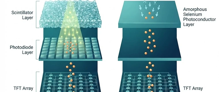
Flat panel detectors have superseded older technologies. These utilize either indirect or direct conversion. Indirect conversion employs a scintillator (e.g., Cesium Iodide) to convert X-rays to light, which a photodiode array then converts to an electrical signal. Direct conversion uses a photoconductor (e.g., Amorphous Selenium) to convert X-rays directly into charge. Direct conversion generally offers superior spatial resolution due to the absence of light spread within the scintillator layer.
| Feature | Indirect Conversion | Direct Conversion |
|---|---|---|
| Intermediate step | Light (scintillator) | None |
| Resolution | Lower (due to light spread) | Higher (direct charge) |
| Efficiency | High (quantum gain) | Moderate |
Exam tip: Distinguish these based on the role of the photoconductor versus the scintillator. Note that indirect conversion is often preferred where lower radiation dose is the primary requirement due to higher quantum gain.
3. Signal-to-Noise Ratio (SNR) and DQE
Detector Quantum Efficiency (DQE) is the definitive measure of how efficiently a detector converts X-ray input into a high-quality output image. A high DQE indicates that the system produces a high signal-to-noise ratio with minimal dose; lower DQE systems require higher radiation doses to achieve the same diagnostic image quality. Modern standards prioritize maintaining high DQE across all digital modalities to comply with current optimization protocols.
Exam tip: A high DQE system allows for lower patient doses without compromising diagnostic accuracy. High DQE is a critical parameter in current procurement specifications.
4. Informatics: PACS, RIS, and Teleradiology
Radiological informatics is a critical component of the physics curriculum. PACS (Picture Archiving and Communication System) manages the storage and retrieval of digital images, while RIS (Radiology Information System) manages workflow, scheduling, and patient data. These systems rely on the DICOM standard to ensure interoperability between vendors, which is essential for effective teleradiology. Recent syllabus updates place increased weight on the physical and engineering components of these infrastructure systems.
Exam tip: DICOM is the universal language of medical imaging, ensuring hardware from different manufacturers can communicate within the teleradiology network.
5. Emerging Trends: PINNs and Room Classification
Physics-Informed Neural Networks (PINNs) are revolutionizing digital image reconstruction by embedding physical laws into machine learning models, improving image correlation, and reducing artifacts. Furthermore, room classifications (Class 1, 2, and 3) have been standardized to define the safety and shielding requirements based on the complexity and invasiveness of the procedural fluoroscopy performed. Adherence to these current classifications is vital for institutional compliance and radiation protection.
Exam tip: PINNs allow for better image reconstruction by constraining neural networks with known physical equations, effectively reducing the noise that would otherwise be amplified by purely data-driven models.
Key Takeaway: The modern digital imaging chain focuses on high DQE detectors, efficient DICOM-based informatics, and the integration of advanced computational methods like PINNs to optimize image quality while minimizing radiation dose in compliance with current SI and safety standards.
Topics: frcr · physics · digital imaging · radiology · informatics